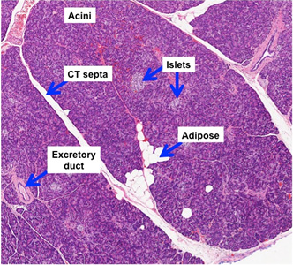
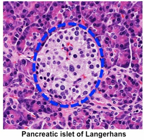
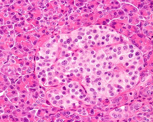
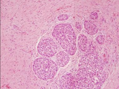
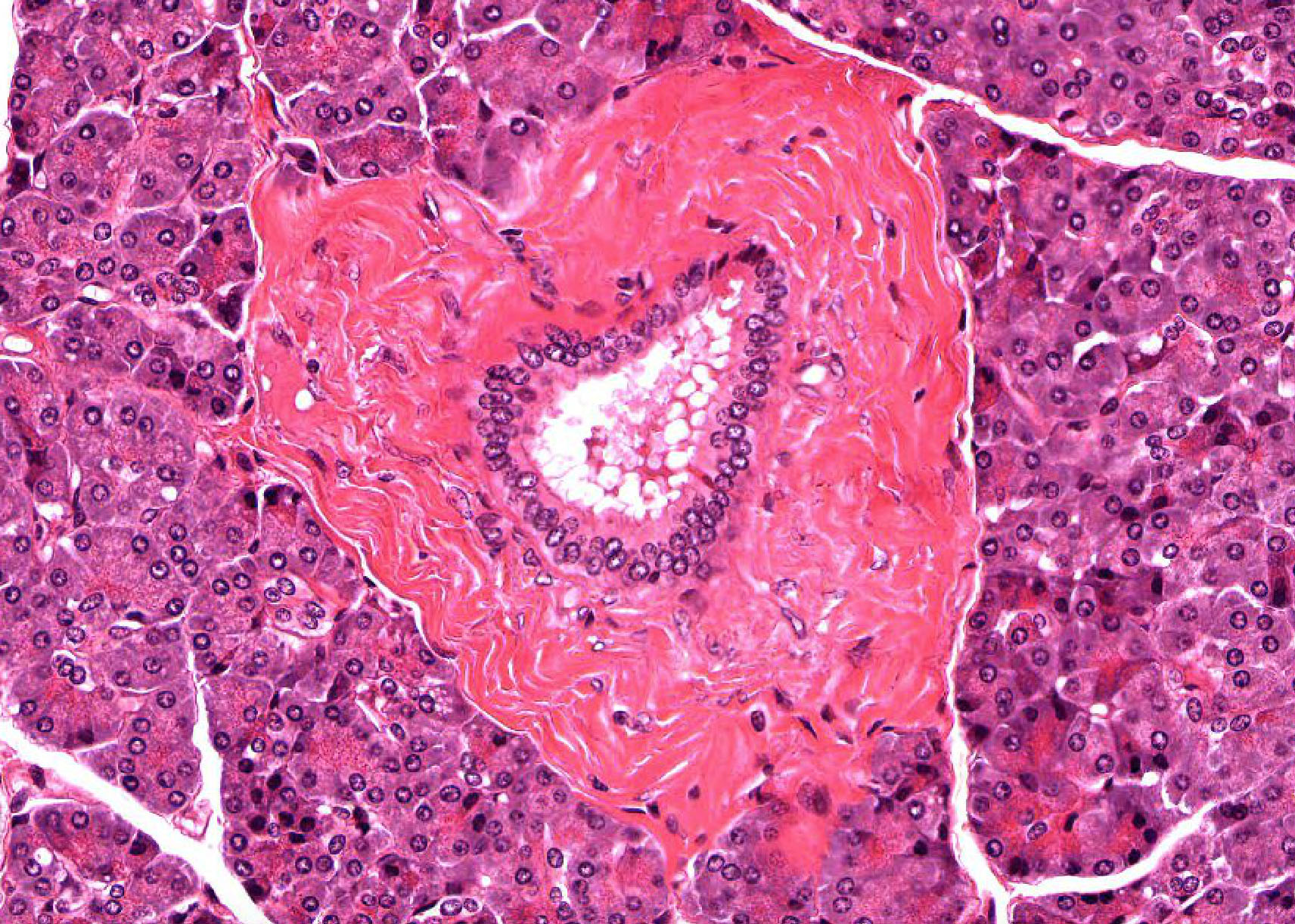
Examine slide 1 at the lowest power and note that most of the section appears purple or bluish This is the parenchyma (or functional tissue) of the exocrine pancreas You will note that the parenchyma is rather indistinctly divided into smaller areas by slits of open space or by pink connective tissue (stroma)Endocrine Pancreas The islets of Langerhans are clumps of secretory cells (up to around 3000) supported by reticulin fibres, and containing numerous fenestrated capillaries There is a delicate capsule around each islet They are paler than the surrounding exocrine cells, as they have less rER These islets do not have an acinar organisation The islet cells are indistinguishable fromThe pancreas of insulinrequiring diabetics becomes progressively less capable of responding to the stimuli used for testing exocrine pancreatic function Frier, Saunders, Wormsley and Bouchier, 1976 and post mortem the whole gland is found to be shrunken and fibrosed Vertiainen, 1944 Furthermore the exocrine cells closest to the islets
Digestive The Histology Guide
Exocrine pancreas under microscope
Exocrine pancreas under microscope-Score to assess acute cellular stress in the human exocrine pancreas This is an open access article under the terms of the Creative Commons Attribution License, which permits use, distribution and reproduction in any visualise islets under a dissecting microscope, further dissected into 1–2mm3 biopsies including visibleIn order to examine the behaviour between the exocrine pancreas under the influence of antiratpancreas immune serum produced in rabbits, a 100 ml immune serum is administered once a week over a maximum 26 week period into Wistarrats by intraperitoneal injection


1
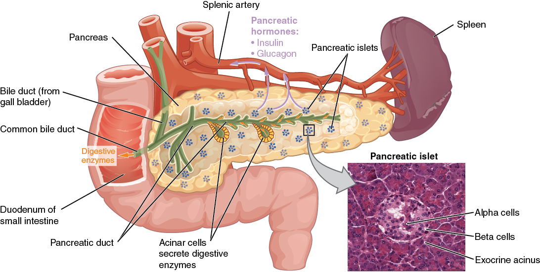
Further, under the microscope, the appearance and arrangement of these carcinoma cells can appear as ductlike (or "adeno") giving the term adenocarcinoma to this most common form of pancreatic cancer About threequarters of exocrine pancreatic cancer arises in the head and neck of the pancreas (the anatomic parts through which the In pancreas histology of animal, you will find the both endocrine and exocrine parts The exocrine part forms the major portion of pancreas and consists of closely packed serous acini with zymogenic cells Again, the endocrine part consists of pancreatic islets of Langerhans which is located within the masses of serous acini The pancreas is both an exocrine accessory digestive organ and a hormone secreting endocrine gland The bulk of the pancreatic tissue is formed by the exocrine component, which consists of many serous pancreatic acini cells These acini synthesize and secrete a variety of enzymes essential to successfully "rest and digest"
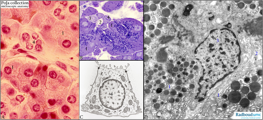
This page has endocrine histology of the pancreas under high powerThe pancreas is a mixed gland meaning it has both endocrine and exocrine tissue When look at the pancreas under the microscope you will see numerous pancreatic acini cells thatIn the exocrine pancreas Given that DHPS catalyzes eIF5A hypusination,1214 and the hypusinated form (eIF5AHYP) functions in mRNA translation,911 we evaluated and uncovered stalled translation elongation concomitant with the differential expression of proteins that influence exocrine pancreas growth and functionThe cells of the pancreatic acini were pyramidal in shape with large spherical and centrally located heterochromatic nuclei The basophilic basal and eosinophilic apical regions seen under the light microscope showed concentrated rough endoplasmic reticulum and homogenous electron dense zymogen granules, respectively, on ultramicroscopy
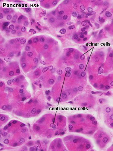
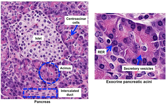
The exocrine portion of the pancreas consists of the pancreatic acini and their ducts The basophilic basal and eosinophilic apical regions seen under theAnswer Acinar cells secrete pancreatic enzymes, centroacinar cells secrete bicarbonate Acinar cells appear more basophilic under the light microscope because of their high RER content and contain many granules under the electron microscopeThese are categorized by looking at the cells under a microscope Doctors use this information to understand the expected growth pattern and speed of the cancer and determine the best course of treatment About 93% of all pancreatic tumors develop in the exocrine component of the pancreas, while only 7% develop in the endocrine component


Exocrine Pancreatic Insufficiency In Dogs Vca Animal Hospital


Digestive The Histology Guide
Using various routine and special techniques and were examined under Leica DM 00 LED microscope During second month of prenatal development, the exocrine pancreas comprised ductules of varying size dispersed within the mesenchyme of both pancreatic primordia As age advanced and the pancreatic primordia fused, theThe architecture of pancreas from organ donors was studied by light and scanning electron microscopy and by wax reconstruction of serial sections It is concluded that the zymogen granulecontaining cells of normal human exocrine pancreas are ar ranged as branching tubules that vary in diameter and curve acutely The endocrine pancreas as it is seen under the microscope The endocrine islets are distributed between the exocrine pancreatic tissue and can have a spherical to ovoid, or strungout, form The threadlike arrangement of the epithelial cells and the dense meshwork of capillaries are apparent


Pancreas Histology Images Stock Photos Vectors Shutterstock


Endocrine Systems Lab
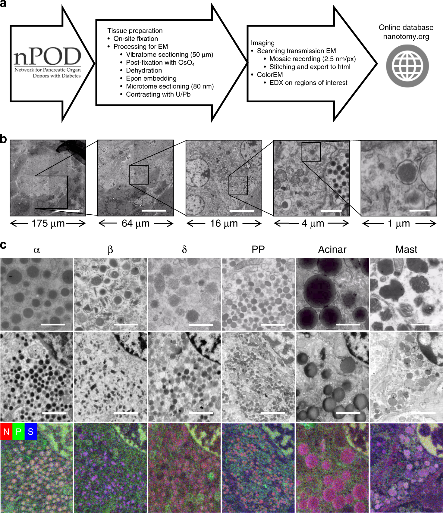
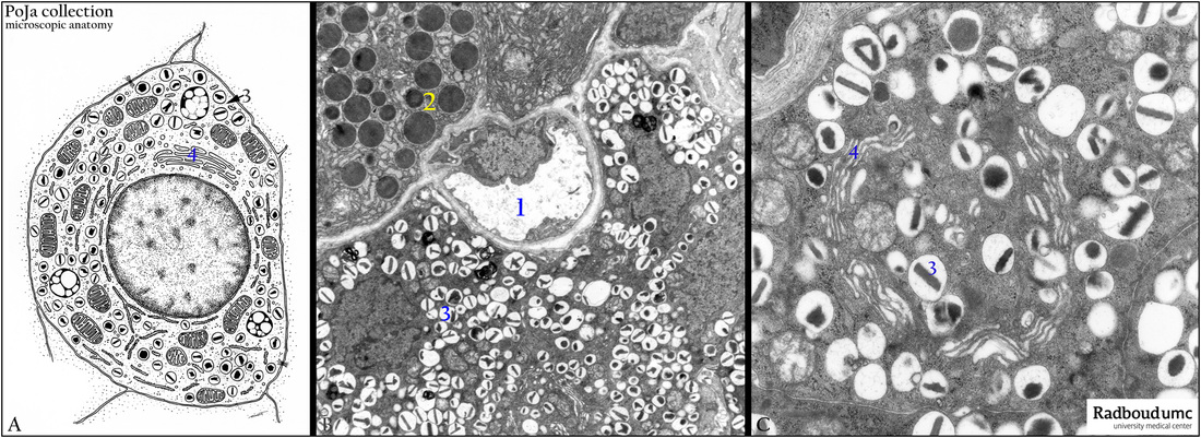
Exocrine pancreatic cancers are the most common type of pancreas cancer Endocrine tumors are more rare and can be benign (not cancerous) or malignant (cancerous) Both tumors can look alike under a microscope, so it isn't always clear if they are really cancer until itPancreatic Insufficiency & Exocrine Pancreas Dysfunction During a consult when there is a problem with digestion I mentally follow a piece of food from the mouth down the digestive tract to the other end and muscle test each anatomical feature as the food passes Wherever the energy is blocked, my fingers will release letting me know what part of the digestive tract needs workCells under the microscope – EM 9 2A The exocrine cell contains a lot of rough ER for protein synthesis The content will be secreted with cytoplasmic vesicles and eventually end up in the a blood b digestive tract 2B The alpha cell produces glucagon, which is visible in the dark vesicles Glucagon secretion ensures that the blood sugar levels


1


Pancreas Histology
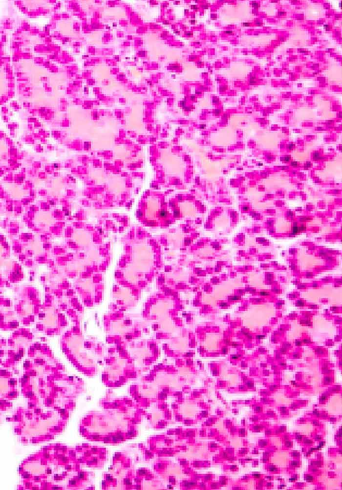
Pancreas 100X The pancreas contains two sets of cells exocrine cells that secrete enzymes via ducts into the intestine, and endocrine cells that secrete hormones into the bloodstream The exocrine cells are small and arranged in spherical secretory units called aciniThe pancreas is made up of mostly exocrine cells, which makes this type of cancer the most common within the pancreas Biopsy — If an imaging test shows an abnormal area, doctors can perform a biopsy and examine tissue under a microscope to learn whether cancer is present Biopsy results help oncologists plan the best treatment for youComparison of an image under a light microscope, a scanning electron microscope and an transmission electron microscope of 212 picometers Applications Structure an exocrine gland 12A1 Structure and function of organelles within an exocrine cell of the pancreas 12A1 Structure and function of organelles within the palisade mesophyll


Pancreas Dr Jastrow S Electron Microscopic Atlas


Human Structure Virtual Microscopy
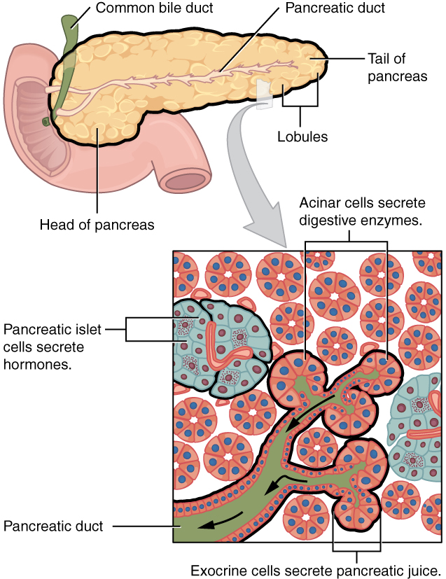
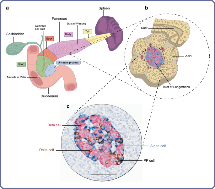
It is possible to distinguish exocrine from endocrine tissue in the pancreas under the light microscope True False True Right after eating a candy bar, insulin secretion would occur True False True Which of the following statement about the pancreas is not true?Pancreas Know the following Islets of Langerhans alpha and beta cells exocrine acinar tissue glucagon produced by alpha cells insulin produced by beta cells Full view of Pancreas Pancreatic islets of Langerhans are small nests of cells, arranged into curvilinear cords, scattered throughout the pancreasThe pancreas contains tissue with an endocrine and exocrine role, and this division is also visible when the pancreas is viewed under a microscope The majority of pancreatic tissue has a digestive role The cells with this role form clusters (Latin acini) around small ducts, and are arranged in lobes that have thin fibrous walls


Chapter 16 Page 3 Histologyolm


Pancreas Anatomy Functions And Diseases Medical Library
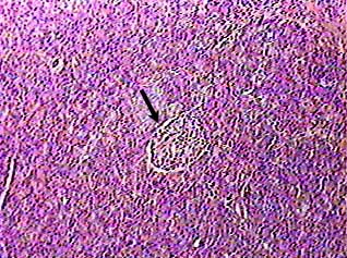
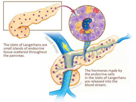
Looking at it under a microscope and it looks 'busy' pancreas is an exocrine organ The exocrine function involves producing digestive Pancreas Pancreas under the stomach Pancreas at low scanning power Duct compared to islets The duct has a very small opening and the islets are labeled with an IBackground During the process of islet isolation, pancreatic enzymes are activated and released, adversely affecting islet survival and function We hypothesize that the exocrine component of pancreases harvested from preweaned juvenile pigs is immature and hence pancreatic tissue from these donors is protected from injury during isolation and prolonged tissue cultureWithin the pancreas, these two functions are performed by different tissues, which can be clearly seen and distinguished from each other when observed under a microscope Most of the cells of the pancreas secrete digestive enzymes (exocrine function) but throughout the organ, there are small sections of cells known as the islets of Langerhans that produce hormones (endocrine


Functional Anatomy Of The Endocrine Pancreas


File Pancreas Histology 002 Jpg Embryology
Lab Content Introduction The term "endocrine" implies secretion into the internal milieu of a multicellular organism In contrast to exocrine tissues, where the secretory products are discharged into the external space the outer surface of the body, mucosal surfaces, duct systems the endocrine organs and cells secrete their products into the vascular system Under a microscope, stained sections of the pancreas reveal two different types of parenchymal tissue2 Lightly staining clusters of cells are called islets of Langerhans, which produce hormones that underlie the endocrine functions of the pancreas Darker staining cells form acini connected to ductsAnatomy of the Pancreas Under a microscope, stained sections of the pancreas reveal two different types of parenchymal tissue Lightstained clusters of cells are called islets of Langerhans These produce hormones that underlie the endocrine functions of the pancreas The darkstained cells form acini that are connected to ducts


Histoquarterly Pancreas Histology Blog


Gross And Microscopic Anatomy Of The Pancreas
HistoQuarterly PANCREAS Working in a histology lab means that I get to see a lot of what our body looks like under the microscope Quarterly I will share with you some of my photos from the microscopic world of our inner space and tell you a little bit about what we're looking at This quarter I have chosen the pancreas because this organ Light microscopic studies of the living acinar pancreas, although limited in number, have revealed valuable information concerning dynamic aspects of microvascular and parenchymal structure and function For example, it has been found that 1) the living organ in anesthetized animals can be imaged with a resolution approaching the limit of the light microscope;A brief review of the normal histology of the pancreas, as presented by the URMC Pathology IT Program


Large Scale Electron Microscopy Database For Human Type 1 Diabetes Nature Communications


Acinar Cells
Exocrine the vast majority of tumors of the pancreas arise in the exocrine part and these cancers look like pancreatic ducts under the microscope These tumors are therefore called "ductal adenocarcinomas," or simply "adenocarcinoma," or even more simply " pancreatic cancer "In the histologic image of an equine pancreas seen below, a single islet is seen in the middle as a large, palestaining cluster of cells All of the surrounding tissue is exocrine Pancreatic exocrine cells are arranged in grapelike clusters called acini (a single one is an acinus)1 Red blood cells Red blood cells are responsible for transporting oxygen throughout the body 2


1


Quantitative Characterization Of The Protein Contents Of The Exocrine Pancreatic Acinar Cell By Soft X Ray Microscopy And Advanced Digital Imaging Methods Unt Digital Library
Human Structure Virtual Microscopy The pancreas is a dual exocrine and endocrine gland The exocrine pancreas is specialized for secretion of digestive enzymes (eg proteinases, lipases, amylases, and nucleases) that are carried by a duct system to the duodenum Small areas of endocrine tissue (the islets of Langerhans) are interspersed amongst the exocrine pancreas andWhat are some features of the exocrine pancreas under a microscope sparse stroma (connective tissue) thin capsule delicate connective tissue septa what is the function of the connective tissue septs in the exocrine pancreas divides parenchyma into distinct lobulesThe pancreatic islets are the exocrine tissue The pancreas produces insulin


Human Structure Virtual Microscopy


Exocrine Pancreas Dr Jastrow S Electron Microscopic Atlas
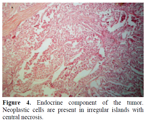
Types of Pancreas Tumors Cancer of the pancreas is not one disease As many as ten different tumor types have been lumped under the umbrella term "cancer of the pancreas", classified as exocrine or endocrine tumorsEach of these tumors has a different appearance when examined with a microscope, some require different treatments, and each carries its own unique prognosis Everything can look strange under a microscope, even the human body Below we have put together a list of images of different parts of the human body under the microscope Some have been increased up to 5,000 times!In the mouse exocrine pancreas cell under the light microscope They are never found in tissue from animals injected with distilled water Although their detailed structure, as seen with the electron microscope, is so varied, they closely resemble the inclusions identified as neutral red granules in earlier work (Byrne, in press)


Liver And Pancreas


Anatomy 15 Virtual Microscopy


Mixed Exocrine Endocrine Tumor Of The Pancreas Insight Medical Publishing


Pancreas Histology Boyar


Pancreatic Histology Exocrine Tissue


Anatomy And Histology Of The Pancreas Pancreapedia


The Ductal System Serving The Exocrine Pancreas Cross Sect Flickr


Exocrine Pancreas Cell Microscopic Photography Macro And Micro Microscopic


Profiling Human Islet Cells With Laser Capture Microdissection


Normal Pancreas Pancreas Org Studying Medicine Histology Slides Medical Laboratory Science


Table Table 1 Definitions For Exocrine Pancreas Tnm Stage 0a Pdq Cancer Information Summaries Ncbi Bookshelf


Pancreas


Vip Reduction In The Pancreas Of F508del Homozygous Cf Mice And Early Signs Of Cystic Fibrosis Related Diabetes Cfrd Journal Of Cystic Fibrosis


Electron Microscopic Study Of Changes In Pancreatic Exocrine Secretory Cells In Both Early And Late Stages Of Hypothyroidism Russian Open Medical Journal


Liver And Pancreas


Regulation Of The Pancreatic Exocrine Differentiation Program And Morphogenesis By Onecut 1 Hnf6 Cellular And Molecular Gastroenterology And Hepatology


Electron Microscopic Study Of Changes In Pancreatic Exocrine Secretory Cells In Both Early And Late Stages Of Hypothyroidism Russian Open Medical Journal


Pancreatic Histology Exocrine Tissue


Implications Of Integrated Pancreatic Microcirculation Crosstalk Between Endocrine And Exocrine Compartments Diabetes


Integrated Pancreatic Blood Flow Bidirectional Microcirculation Between Endocrine And Exocrine Pancreas Diabetes


Dictionary Normal Pancreas The Human Protein Atlas
:watermark(/images/watermark_5000_10percent.png,0,0,0):watermark(/images/logo_url.png,-10,-10,0):format(jpeg)/images/overview_image/1881/CPsIC67xuz6pLt6JhAM6Wg_pancreas-histology_english.jpg)


Pancreas Histology Exocrine Endocrine Parts Function Kenhub


The Healthy Exocrine Pancreas Contains Preproinsulin Specific Cd8 T Cells That Attack Islets In Type 1 Diabetes


Pancreas Physiology Intechopen


Morphological Histological And Ultrastructural Studies On The Exocrine Pancreas Of Goose Sciencedirect


Solved 3 Pancreas 1 Pancreatic Acini Exocrine Portion 2 Chegg Com


Objective 4 4c Cytoplasmic Organelles Secretory Granules Em


Exocrine Pancreas Biochemistry Flashcards Draw It To Know It


Distribution Of Exocrine Portion Of The Pancreas A The Pancreas Is Download Scientific Diagram


Anatomy And Histology Of The Pancreas Pancreapedia


The Roles Of Calcium And Atp In The Physiology And Pathology Of The Exocrine Pancreas Physiological Reviews


The Endocrine And Exocrine Pancreas Cross Section Human P Flickr


Organisation Of The Human Pancreas In Health And In Diabetes Springerlink


B Cells In Islet Of Langerhans In The Exocrine Pancreas Dog Poja Collection Microscopic Anatomy


Pancreas 400x


Types Of Pancreatic Cancer Pancreatic Cancer Action Network


Human Structure Virtual Microscopy


What Are Pancreatic Causes Of Exocrine Pancreatic Insufficiency Epi


Chapter 16 Page 3 Histologyolm


Loss Of The Ciliary Protein Chibby1 In Mice Leads To Exocrine Pancreatic Degeneration And Pancreatitis Scientific Reports


Pancreas Histology


Scanning Electron Microscopy Of The Rat Exocrine Pancreas Semantic Scholar


Pancreas Wikipedia


Microscopic Section Of Exocrine Pancreatic Adenocarcinoma Invasion Of Download Scientific Diagram


Exocrine Pancreas An Overview Sciencedirect Topics
/exocrine-pancreatic-insufficiency-4177936-821-618578bea845406fa96c78167aff8657.png)


Exocrine Pancreatic Insufficiency Symptoms Causes And Diagnosis


A Microscopic Section Of Exocrine Pancreatic Adenocarcinoma The Download Scientific Diagram


Endocrine Pathology
:watermark(/images/watermark_only.png,0,0,0):watermark(/images/logo_url.png,-10,-10,0):format(jpeg)/images/anatomy_term/body-of-pancreas-3/mbuhIx2qC3HVaYNH69w_Body_of_Pancreas.png)


Pancreas Histology Exocrine Endocrine Parts Function Kenhub


Electron Microscopic Study Of Changes In Pancreatic Exocrine Secretory Cells In Both Early And Late Stages Of Hypothyroidism Russian Open Medical Journal


Liver And Pancreas


Histology Laboratory Manual


Chapter 16 Page 3 Histologyolm


Scanning Electron Microscopy Of The Rat Exocrine Pancreas Semantic Scholar


Pancreas Anatomy Functions And Diseases Medical Library


Pancreas Function Pancreatic Cancer Johns Hopkins Pathology


Exocrine Pancreas Sciencedirect


Pancreatic Histology Exocrine Tissue
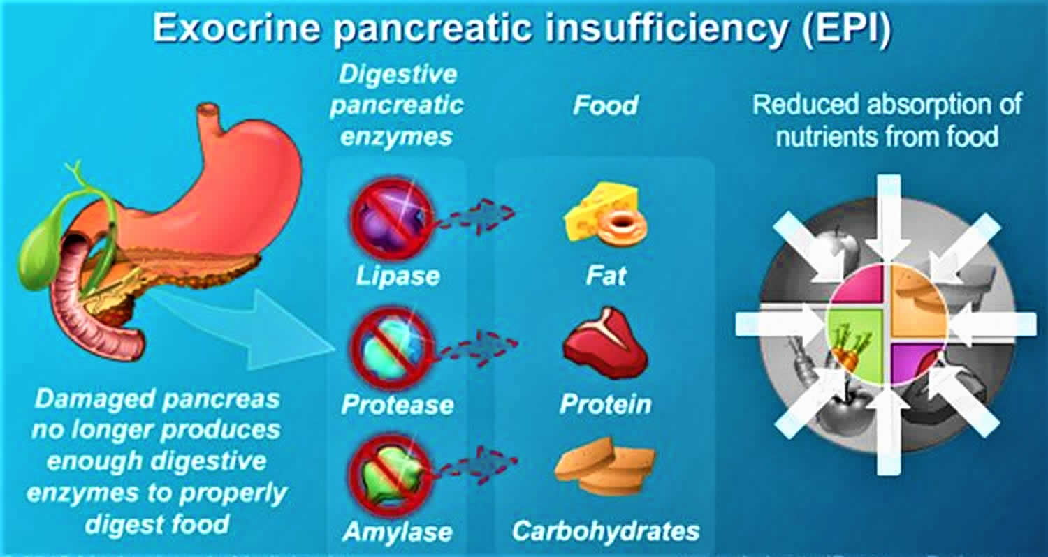


Exocrine Pancreatic Insufficiency Causes Symptoms Diagnosis Treatment


Pancreas Wikipedia
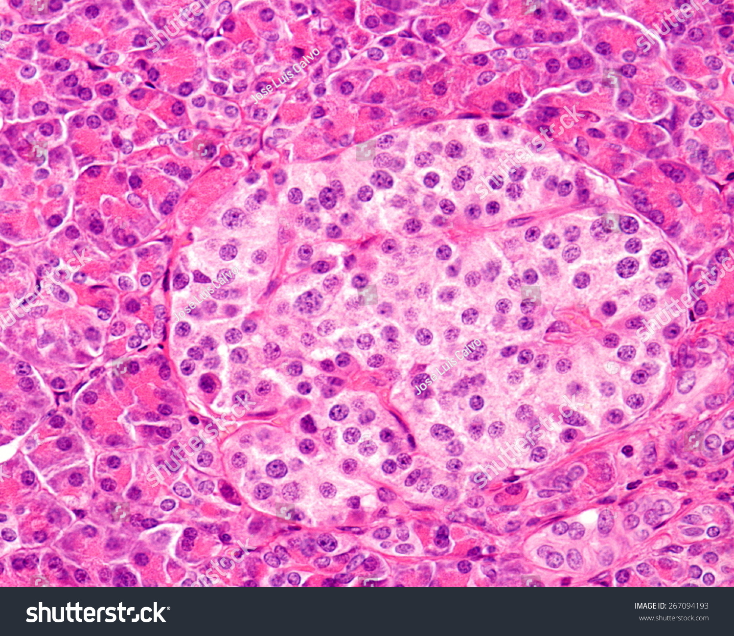


High Magnification Of A Human Islet Of Royalty Free Stock Photo Avopix Com
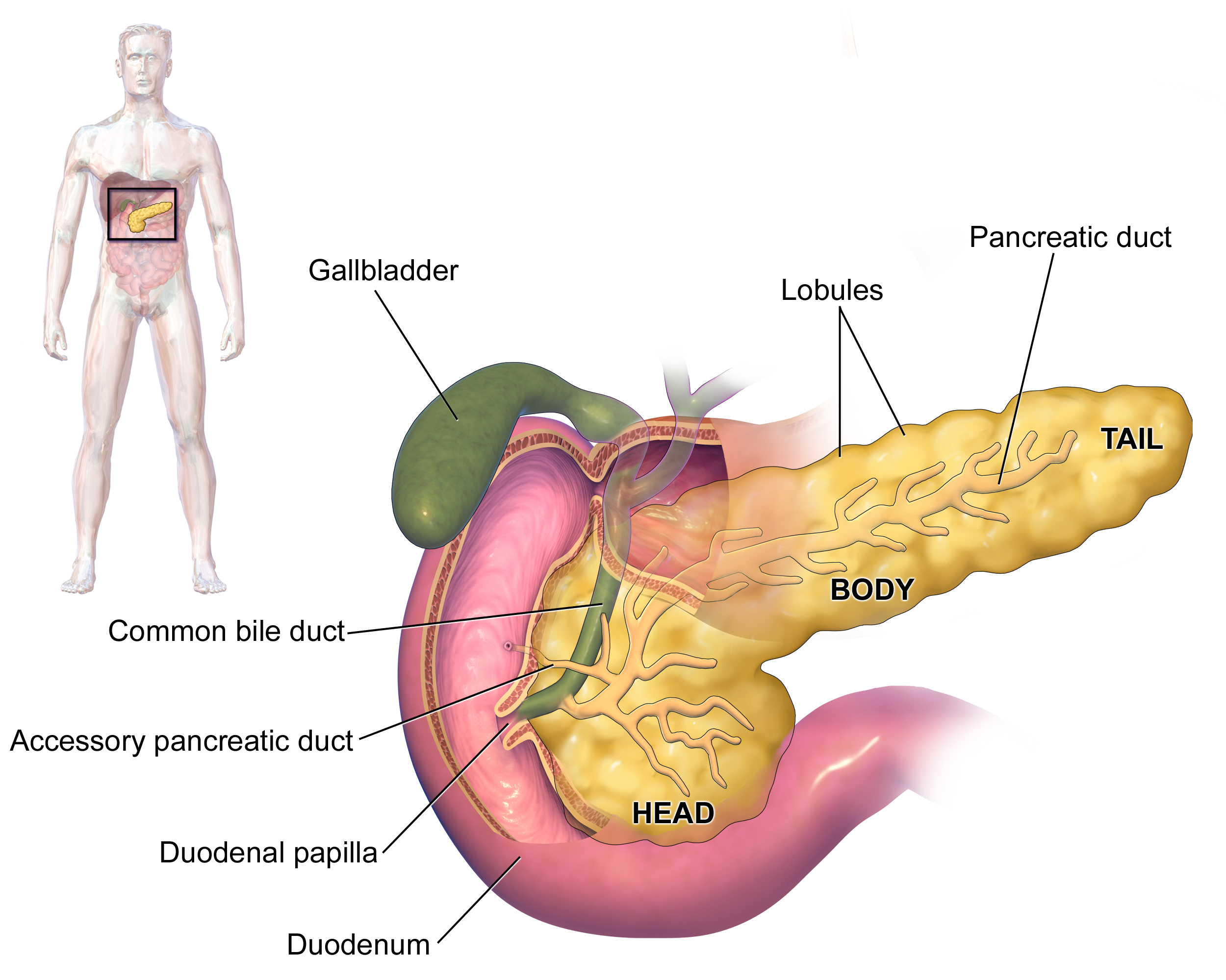


Pancreas Wikipedia
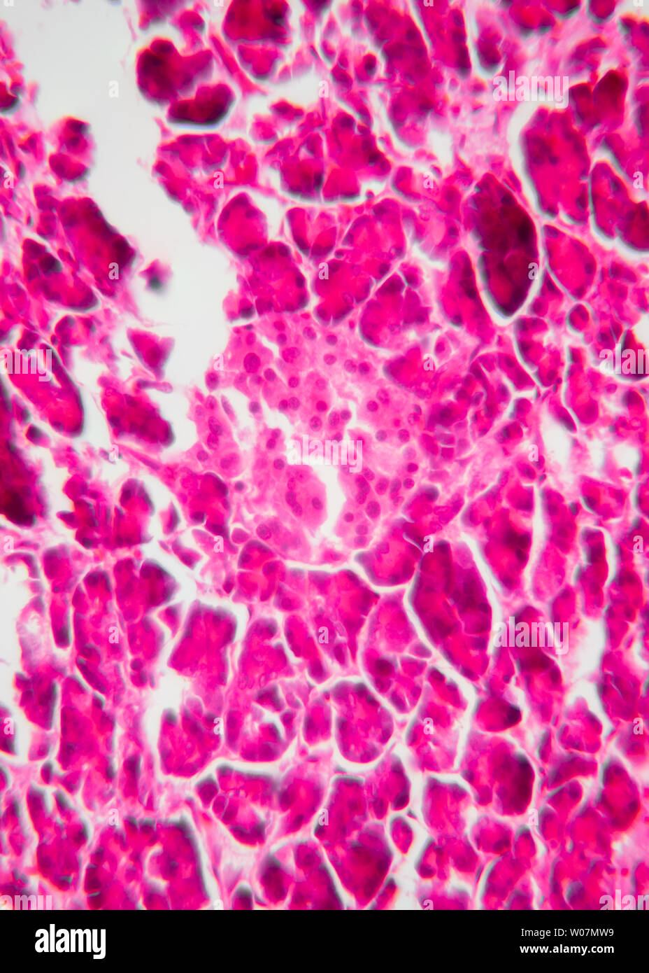


The Endocrine Part Of The Pancreas Islet And The Exocrine Part Magnified By An Optical Microscope 400 Times Shows The Islet Stock Photo Alamy


Liver And Pancreas


Exocrine And Endocrine Pancreas Light Micrograph Stock Image C036 1331 Science Photo Library


Pancreas Histology


Labeled


Histoquarterly Pancreas Histology Blog


Histology Of The Pancreas Endocrine And Exocrine Youtube


Histoquarterly Pancreas Histology Blog


Intercalated Duct Of Exocrine Pancreas Human Rabbit Poja Collection Microscopic Anatomy


Electron Micrograph Of Acinar Cells Of The Exocrine Pancreas Download Scientific Diagram


P2ry1 Alk3 Expressing Cells Within The Adult Human Exocrine Pancreas Are Bmp 7 Expandable And Exhibit Progenitor Like Characteristics Cell Reports


Liver Gall Bladder Pancreas


Pancreas Histology


Organisation Of The Human Pancreas In Health And In Diabetes Springerlink
:watermark(/images/watermark_only.png,0,0,0):watermark(/images/logo_url.png,-10,-10,0):format(jpeg)/images/anatomy_term/pancreatic-acinar-cells/86g1HQPABGT8iQy0zPdrpg_Exocrine_cell_of_pancreas.png)


Pancreas Histology Exocrine Endocrine Parts Function Kenhub


Pancreas Histology Identifying Features With Labeled Slide Images Anatomylearner The Place To Learn Veterinary Anatomy Online


Anatomy 15 Virtual Microscopy


Pancreas Function Pancreatic Cancer Johns Hopkins Pathology


Exocrine Cell Of Pancreas Electron Micrograph Labelling Diagram Quizlet


Anatomy And Histology Of The Pancreas Pancreapedia


Men1 Maintains Exocrine Pancreas Homeostasis In Response To Inflammation And Oncogenic Stress Pnas


Color Scanning Electron Micrograph Of An Acinar Exocrine Pancreatic Cell Acinar Cells Produce And Excrete Digestive Enzymes To The Small Intestine Via The Pancreatic Ducts Stock Photo Offset


0 件のコメント:
コメントを投稿